
ANATOMY & PHYSIOLOGY 20132014 Urinary System
Lab 9: Anatomy of the Urinary System. A&P Lab Manual. Lab 9: Anatomy of the Urinary System. Atlas: Urinary System. Additional Activities: Lab 9. Models of the Urinary System - Blank. Models of the Urinary System - Labeling Activity. Practice Quiz. Urinary Anatomy Practice Quiz . Lab Model Videos.

Diagram Of Urinary System With Labels
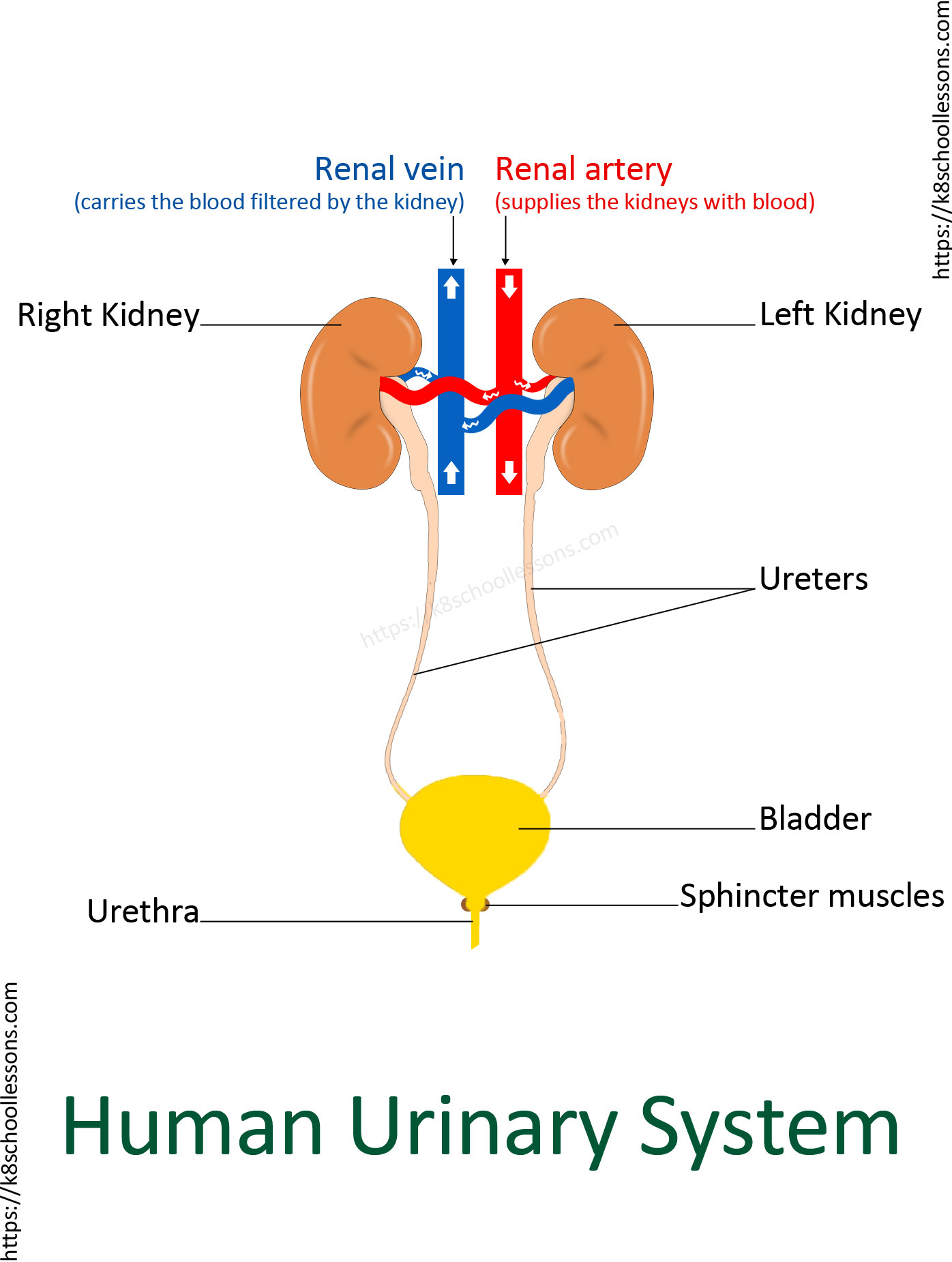
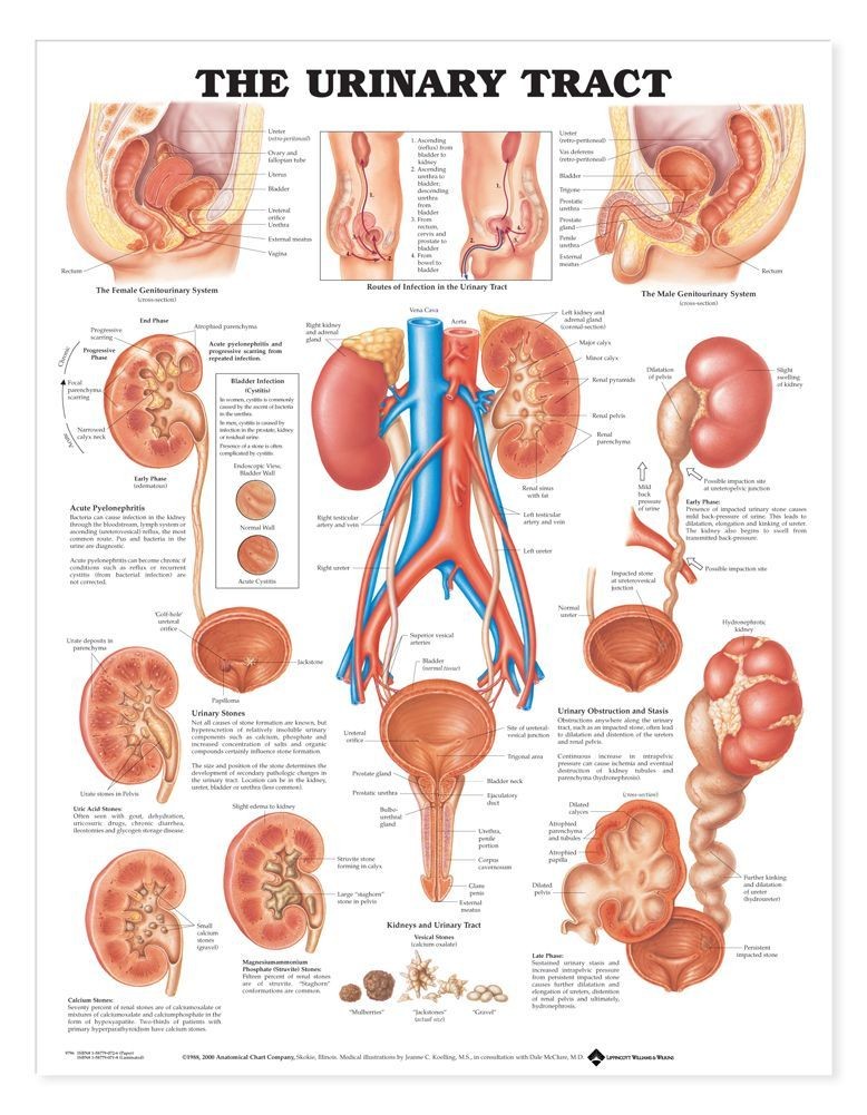
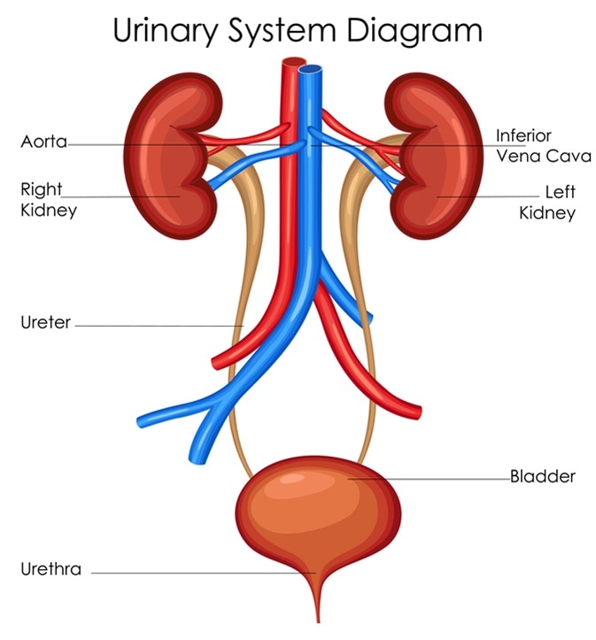
Bladder. This triangle-shaped, hollow organ is located in the lower abdomen. It is held in place by ligaments that are attached to other organs and the pelvic bones. The bladder's walls relax and expand to store urine, and contract and flatten to empty urine through the urethra.
Urinary System for Kids Human Urinary System Human Body Facts
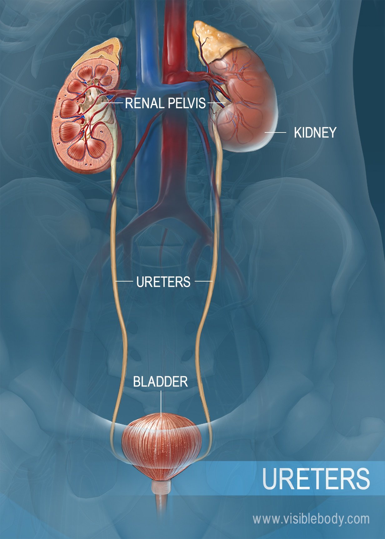
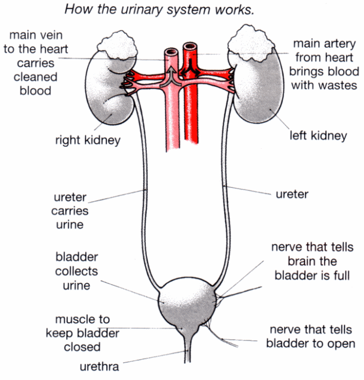
The uterus is also shown. Anatomy of the female urinary system showing the kidneys, ureters, bladder, and urethra. Urine is made in the renal tubules and collects in the renal pelvis of each kidney. The urine flows from the kidneys through the ureters to the bladder. The urine is stored in the bladder until it leaves the body through the urethra.
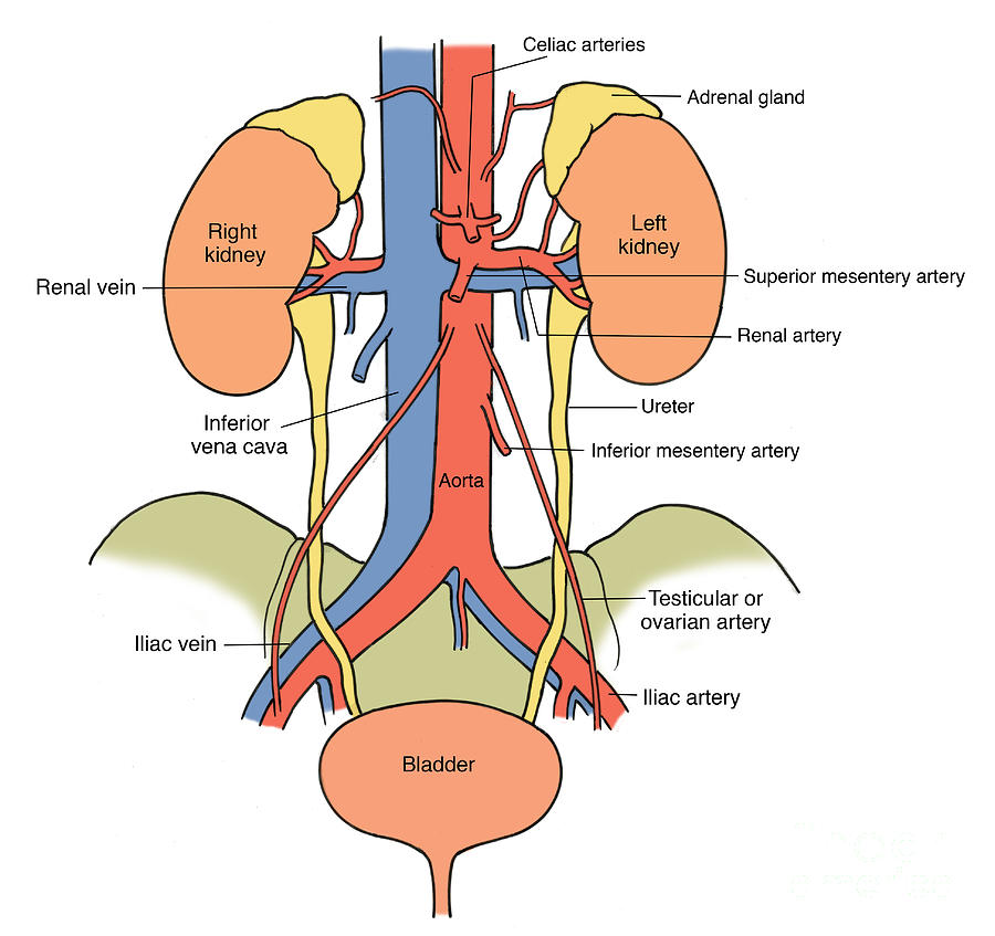
Illustration Of Urinary System Photograph by Science Source
NEET 2023 Answer Key Human Urinary System Diagram Humans get rid of wastes from the body through the urinary system. The urinary system is functional in turning toxic substances into the urine, storing and carrying urine, and safely eliminating it from the body.
The Urinary Tract System Chart MedWest Medical Supplies
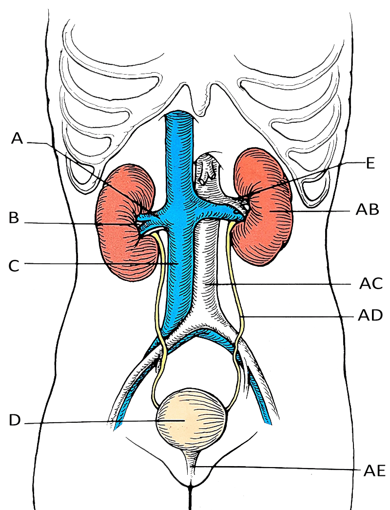
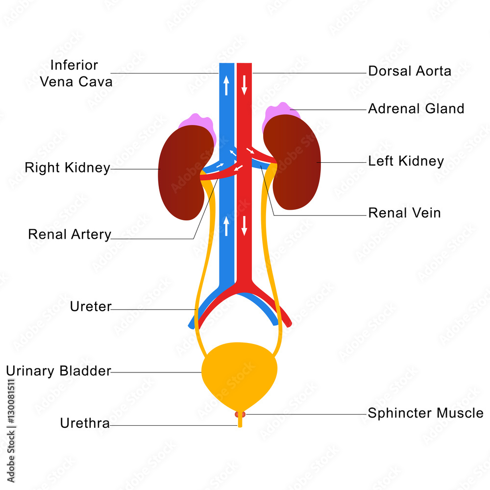
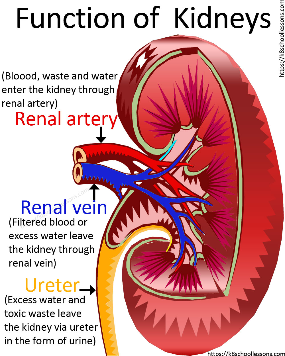
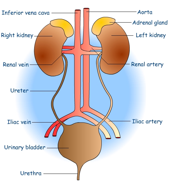
urinary system that removes nitrogenous wastes from the body. The urinary system is also responsible for maintaining the electrolyte, acid-base, and fluid balances of the blood and is thus a major, if not the major, homeostatic organ system of the body. The primary organs in the urinary system are the paired kidneys (Figure 1). To properly do.
Label the Urinary System
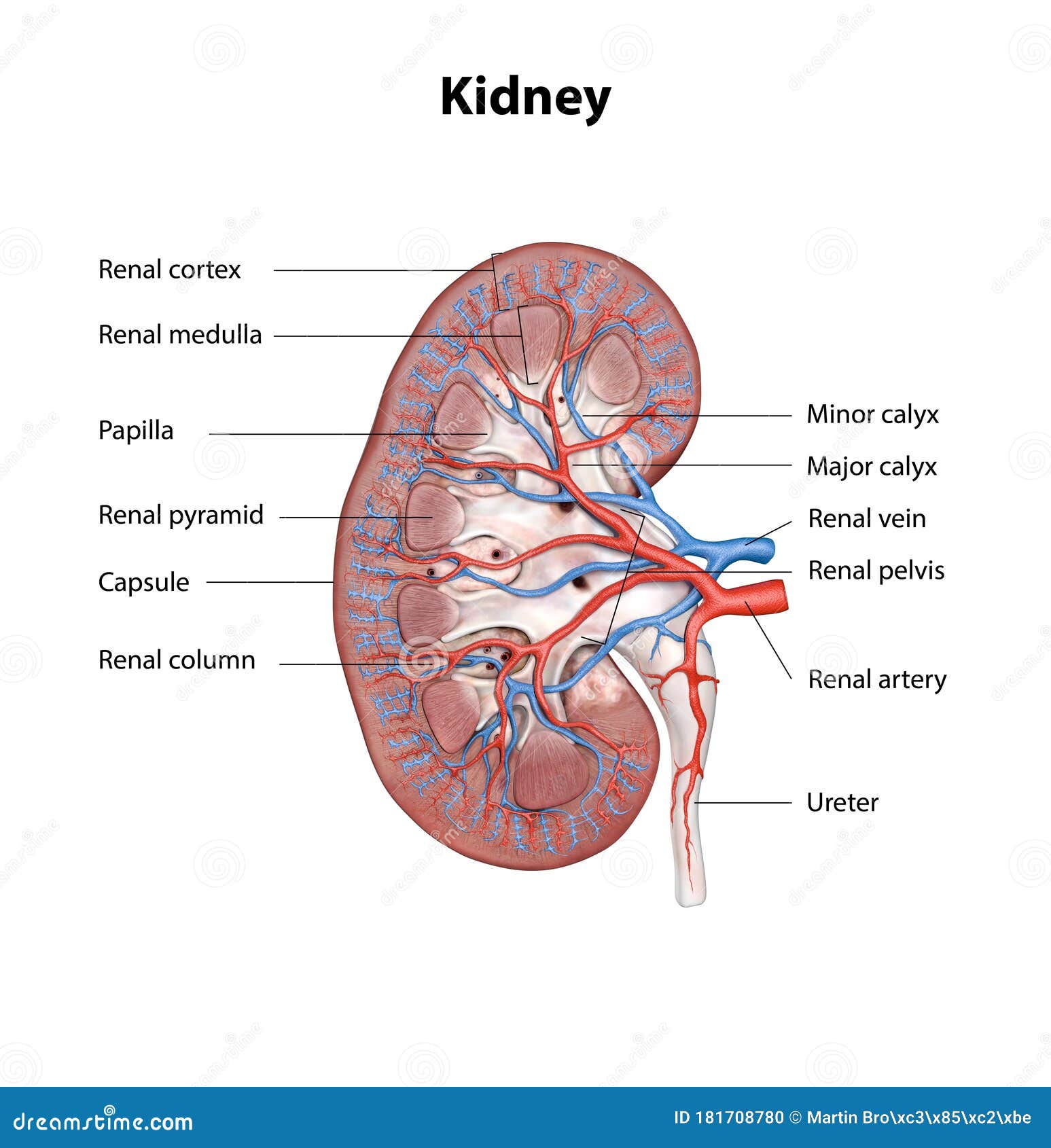
1/3 Synonyms: none The kidneys are bilateral organs placed retroperitoneally in the upper left and right abdominal quadrants and are part of the urinary system. Their shape resembles a bean, where we can describe the superior and inferior poles, as well as the major convexity pointed laterally, and the minor concavity pointed medially.
Urinary System Structures
April 5, 2021 in Anatomy, Worksheets by Shannan Muskopf anatomy, kidney, label, learn, nephron, practice, urinary Students practice labeling the urinary system with this drag and drop activity. Three slides have detailed images of the kidneys, ureters, and nephrons.
Human urinary system labelled diagram Stock Vector Adobe Stock
View All Diagram External Internal Breast Anatomy Functions Female anatomy includes the internal and external structures of the reproductive and urinary systems. Reproductive anatomy plays a role in sexual pleasure, getting pregnant, and breastfeeding. The urinary system helps rid the body of toxins through urination (peeing).
Urinary System for Kids Human Urinary System Human Body Facts
The primary structures of the urinary system include the kidneys, ureters, bladder, and urethra. Learn about the complex role of the kidneys, how urine drains into the ureters, and just how much the adult bladder can hold. Learn the main differences between the female and male urethra. Read Post.
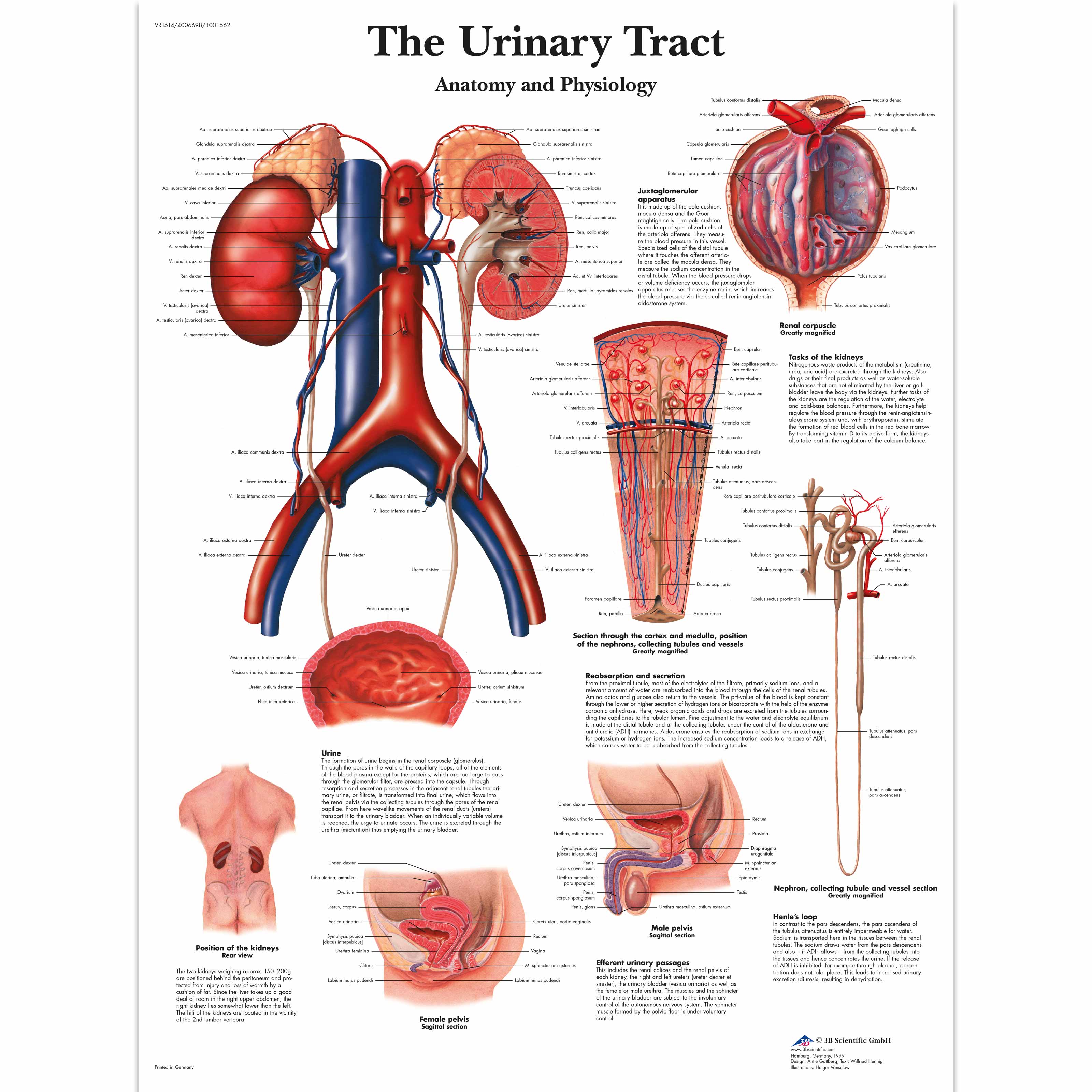
The Urinary Tract Anatomy and Physiology 1001562 3B Scientific VR1514L Urinary system
Health Library / Body Systems & Organs / Urinary System Urinary System The urinary system includes your kidneys, ureters, bladder and urethra. This system filters your blood, removing waste and excess water. This waste becomes pee. The most common urinary issues are bladder infections and urinary tract infections (UTIs).

Structure and function of urinary system Vector Image
The urinary bladder and urethra are pelvic urinary organs whose respective functions are to store and expel urine outside of the body in the act of micturition (urination). As is the case with most of the pelvic viscera, there are differences between male and female anatomy of the urinary bladder and urethra. In our entire urinary system series, the urinary bladder and urethra represent the.
Structure of the Bladder
Ren 1/5 Synonyms: none The urinary system consists of 4 major organs; the kidneys, ureters, urinary bladder and the urethra. Together these organs act to filter blood, remove waste products, create urine and transport urine out from the body.

The Urinary System 2600 Anatomical Parts & Charts
Some of the original labels from the wikimedia file were removed to make the diagram simpler and more specific to just the urinary system.. Diaper Drama Urinary System - Label the Kidney and Nephron Muscles Labeling Neuroglia Labeling with Google Slides. Posted . May 3, 2020. in .
urinary system diagram
Kidney cross section This cutaway shows the kidney's main layers, the cortex and the medulla, which form segments known as renal pyramids. The renal artery and vein circulate huge amounts of blood - about 2 1/2 pints/min at rest, which is up to one-quarter of the heart's total output.
urinary system parts
Urinary System Diagram. Image Credit: Vecton / Shutterstock. Ureter. The ureters are tubes which expel urine from the kidneys. Within the human body there are two ureters, one connected to each.
Human Kidney Cross Section, Scientific Background, Anatomy, Urinary System with Main Parts
Last updated on September 21, 2023 The urinary system, also known as the renal system, is an essential part of the body responsible for the production, storage, and elimination of urine.
