
Account Suspended Heart diagram, Human heart diagram, Biology worksheet
How to Draw Human Heart Diagram Drawing / easy way - Step by step - YouTube © 2023 Google LLC Hi friends,very warm welcome to all from the bottom of my Heart,Your heart's main function.

Labeled Pictures Of the Heart Lovely Simple Human Heart Diagram for Kids Human heart diagram
Diagram of Heart The human heart is the most crucial organ of the human body. It pumps blood from the heart to different parts of the body and back to the heart. The most common heart attack symptoms or warning signs are chest pain, breathlessness, nausea, sweating etc.

labled heart diagram
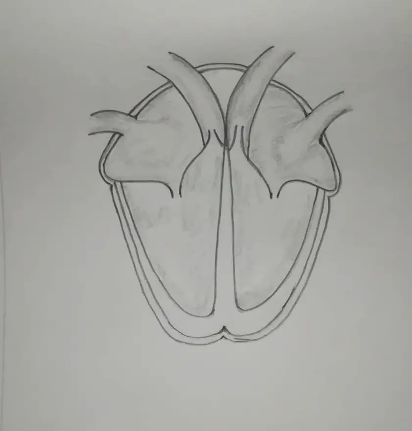
Drawing a human heart is easier than you may think. With our step-by-step guide, you'll be sketching and shading a realistic heart in no time. Plus, you may just learn something new along the way. So, grab your pencil and sketchbook because we've got a heart to draw! Method 1 Sketching the Heart Download Article 1

Free Blank Heart Diagram, Download Free Blank Heart Diagram png images, Free ClipArts on Clipart
In this blog post, we'll provide an easy and simple diagram of the human heart to help you understand how it works. The Anatomy of the Human Heart. The human heart is a muscular organ that pumps blood throughout the body. It is located in the chest, slightly to the left of the sternum. The heart is divided into four chambers: the right atrium.

Heart diagram (annotated) Openclipart
The heart blood flow diagram (flowchart) given below will help you to understand the pathway of blood through the heart.Initial five points denotes impure or deoxygenated blood and the last five points denotes pure or oxygenated blood. 1.Different Parts of the Body. ↓. 2.Major Veins.

Printable Human Heart Diagram Printable Word Searches
The human heart is located within the thoracic cavity, medially between the lungs in the space known as the mediastinum. Figure 19.2 shows the position of the heart within the thoracic cavity. Within the mediastinum, the heart is separated from the other mediastinal structures by a tough membrane known as the pericardium, or pericardial sac.
Structure of the Heart YouTube
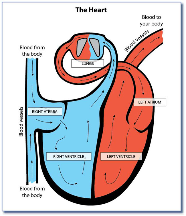
The heart, nestled between the lungs and protected by the rib cage, serves as a powerful pump ensuring blood flow throughout the body. This systemic flow delivers oxygen and nutrients to cells and removes waste. Additionally, the heart manages pulmonary flow, sending blood to the lungs for oxygenation before distributing it to the body.

FileHeart Diagramen.svg Wikipedia, The Free Encyclopedia Cliparts.co
Your heart is the primary organ of your circulatory system. It pumps blood throughout your body, controls your heart rate and maintains blood pressure. Your heart is a bit like a house. It has walls, rooms, doors, plumbing and an electrical system. All the parts of your heart work together to keep blood flowing and send nutrients to your other.
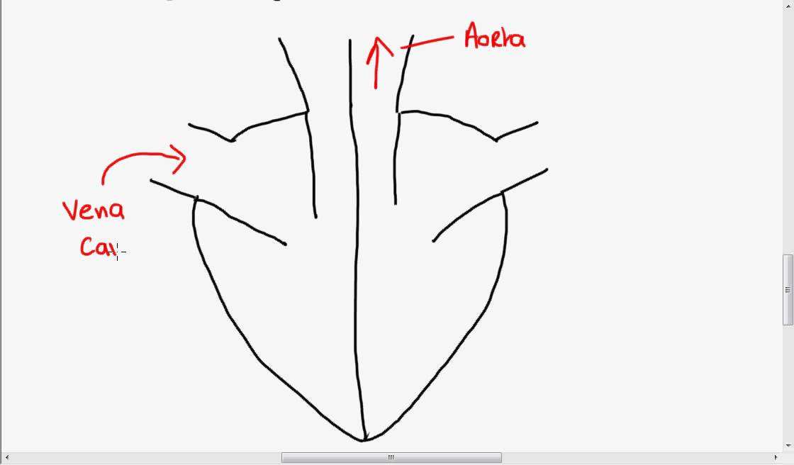
How To Draw Human Heart Diagram
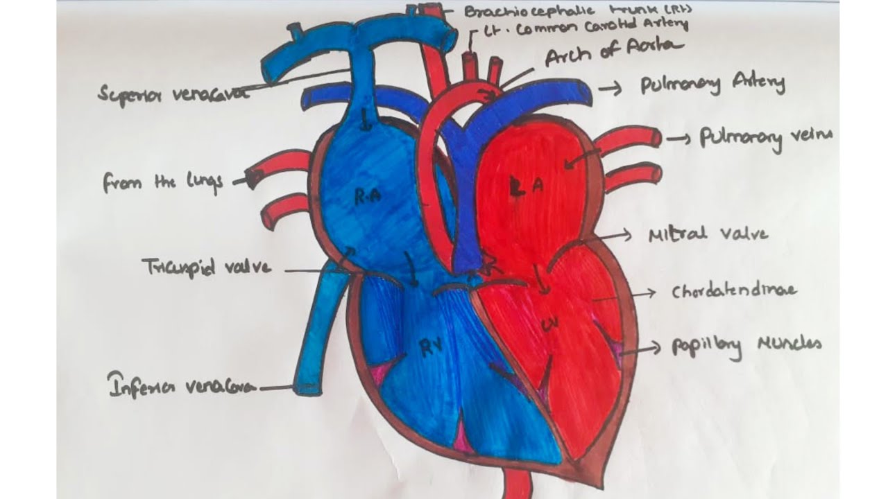
The human heart is primarily comprised of four chambers. The two upper chambers are called the atria, the remaining two lower chambers are the ventricles. The right and left sides of the heart are separated by a muscle called the "septum.". Both sides work together to efficiently circulate the blood.

Come Disegnare la Struttura Interna del Cuore
The easiest way to draw a heart diagram is using EdrawMax by simply choosing a template, then export it as any formats you prefer and attach in your science report, presentation, assignment and so on. Try EdrawMax Now 1. What Does the Heart Look Like The heart is a muscle.

Heart Labeling Worksheet
2. Next the blood enters the lungs and performs gas exchange, the blood now has high O2 and goes to the pulmonary vein back towards the heart. 3. This newly oxygenated blood goes through the left side and out the aorta, the main artery going from the heart to the body.

diagram of human heart easy Online Sale, UP TO 58 OFF
The heart is made of three layers of tissue. Endocardium is the thin inner lining of the heart chambers and also forms the surface of the valves.; Myocardium is the thick middle layer of muscle that allows your heart chambers to contract and relax to pump blood to your body.; Pericardium is the sac that surrounds your heart. Made of thin layers of tissue, it holds the heart in place and.
Update more than 155 human heart drawing simple best vietkidsiq.edu.vn
How to Draw Human Heart Step by Step!Our Website: http://bit.ly/2KBC0l1Android App: https://bit.ly/3k48zdKCBSE Class 10 Courses: https://bit.ly/363U55VCBSE C.

5th Grade Mrs.Asoklis/HEART
How to draw human heart step by step, easy trick to draw human heart, how to draw the diagram of human heart, easily drawing diagram of human heart for cla.

Pin on Cardio
+ Show all Heart anatomy The heart has five surfaces: base (posterior), diaphragmatic (inferior), sternocostal (anterior), and left and right pulmonary surfaces. It also has several margins: right, left, superior, and inferior: The right margin is the small section of the right atrium that extends between the superior and inferior vena cava .
Health & PE 06
Function and anatomy of the heart made easy using labeled diagrams of cardiac structures and blood flow through the atria, ventricles, valves, aorta, pulmonary arteries veins, superior inferior vena cava, and chambers. Includes an exercise, review worksheet, quiz, and model drawing of an anterior view (frontal section) of the heart in order to.