
The Cervical Spinal Cord and Origin of the Anterior Spinal Artery. The... Download Scientific
Vena vertebralis anterior Quick Facts Origin Course Tributaries Structures Drained Quick Facts Origin: Around the transverse processes of the upper cervical vertebrae. Course: Descends in the neck to drain into the vertebral vein. Tributaries: Anterior external vertebral venous plexus. Drainage: Vertebral column. Complete Anatomy

Vascular anatomy
Description The vertebral venous plexuses are the network of veins that surround the vertebral column along its entire length. It is a nonuniform network of vessels located inside the vertebral column and immediately outside the vertebrae, primarily concentrated in anterior and posterior locations.
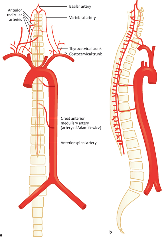
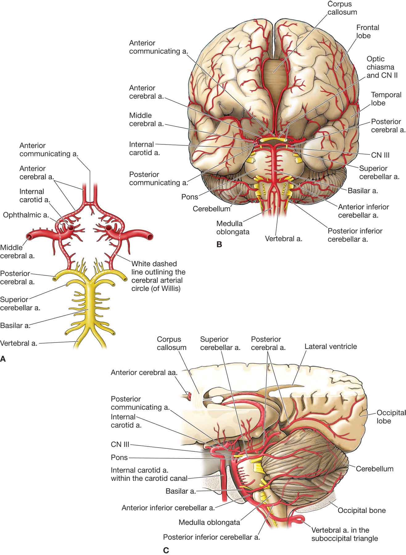
Arteries of the Spinal Cord Radiology Key
The lumbar veins are four pairs of blood vessels that drain the lumbar segments of the spinal cord, posterolateral abdominal wall and lumbar structures of the back. They usually empty into the inferior vena cava, but they can also drain into the ascending lumbar, azygos, renal or other lumbar veins.

vertebral artery course Vertebral artery, Medical anatomy, Medical knowledge
The vertebral vein descends in the foramina transversaria of the upper six cervical vertebrae and forms a plexus around the vertebral artery. After emerging from the foramen transversarium of the sixth cervical vertebra, the vertebral vein descends anterolateral to the vertebral artery. It passes behind the internal jugular vein and anterior to.
Vertebral Artery Anatomy Anatomical Charts & Posters
The vertebral vein (Latin: vena vertebralis) is a venous blood vessel that is formed by numerous small tributaries arising from the internal vertebral plexuses. The vertebral vein collects venous blood mainly from the upper deep muscles of the back. Vertebral vein by Anatomy.app
Vertebral Artery Art Print by Asklepios Medical Atlas
The blood supply to the vertebral canal is critical especially from the context of surgical and clinical considerations. The spinal cord located within the vertebral canal allows for a neuronal connection between the brain and the rest of the body and thus the blood supply to this structure is of vital importance. In this review, we go over the blood supply as well as additional information.

Veins of Vertebral Column Vertebral Veins Anatomy Superior bulb of right internal jugular vein
Spinal Vascular Malformations. This section is intended as an introduction to the (daunting) topic of spinal vascular anatomy. As interventionalists, we usually deal with two broad types of spinal issues — various vascular malformations and tumors. In the former group, we are mainly concerned with fistulas (dural, pial, cord) — not because.

Vertebral Artery Anatomy Anatomical Charts & Posters
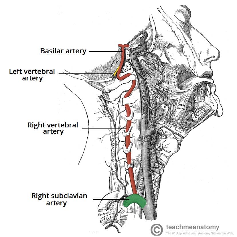
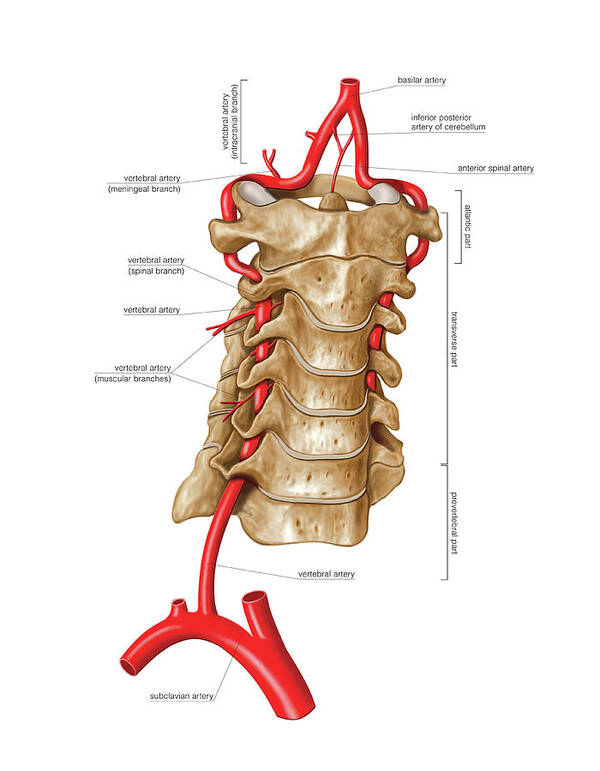
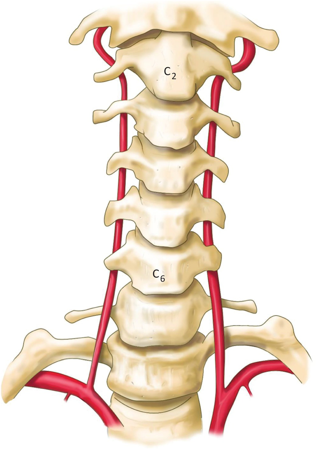
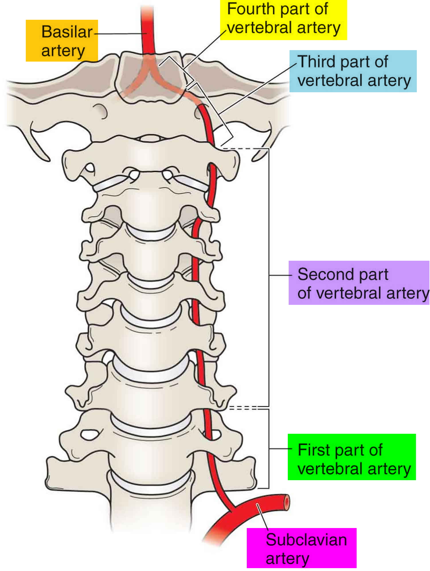
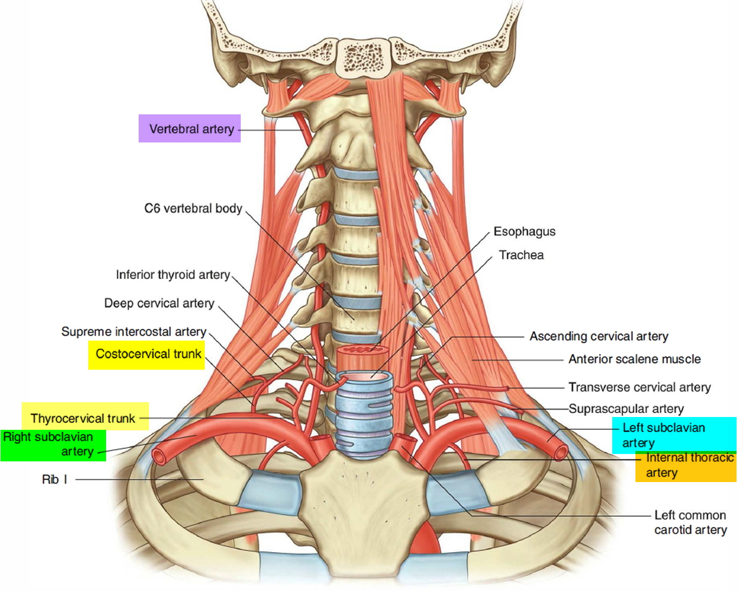
Vertebral Arteries (VA): 4 Segments V1 - Extraosseous segment Origin from each subclavian artery, coursing superiorly to enter the C6 transverse foramen (80-90%) Variations Origin of the left vertebral artery from the aortic arch between the left common carotid artery and left subclavian artery has been described in 2-6% of cases
Vertebral Artery Anatomy Anatomical Charts & Posters
a -centripetal network of veins, predominantly draining the gray matter into: b - central (sulcal) veins of the intrinsic system; c - peripheral (radial, a.k.a. marginal) centrifugal veins of the intrinsic system; d - venous anastomosis between the centripetal and centrifugal systems; e - anterior (ventral) median vein; f - posterior (dorsal) me.
Vertebral Artery Segments, Stenosis and Artery Dissection Symptoms
Structure and Function The spinal venous system consists of the intrinsic, extrinsic, and extradural systems. During IVC occlusion, the spinal venous system can serve as a collateral drainage system due to its connection with both pelvic and cranial veins. [1]

Veins of the Vertebral Column (preview) Human Anatomy Kenhub YouTube
The vertebral vein is a paired blood vessel that is found on either side of the neck. As a single vessel, it is formed at the level of the sixth cervical vertebra (C6). Its initial part is a venous plexus that goes through the transverse foramina of the cervical vertebrae. Check it out
Vertebral Artery Segments, Stenosis and Artery Dissection Symptoms
Starting at vertebral levels T12-L2, the azygos vein travels posterior to the right root of the lung (T5-T6) and arches superiorly over the root of the lung, emptying into the SVC. Sometimes, the azygos vein may travel higher to drain into the brachiocephalic or right subclavian vein.
Vertebral Artery Musculoskeletal Key
Anatomy of Spinal Venous Drainage for the Neurointerventionalist: From Puncture Site to Intervertebral Foramen N. Borg, J. Cutsforth-Gregory, S. Oushy, T. Huynh, L.E. Savastano, H.J. Cloft, G. Lanzino and W. Brinjikji American Journal of Neuroradiology January 2022, DOI: https://doi.org/10.3174/ajnr.A7409 Article Figures & Data Info & Metrics

Vertebral Artery Anatomy Anatomical Charts & Posters
The vertebral vein is a paired vessel found in the transverse foramina of the cervical vertebrae on either side of the neck. It arises at the level of the sixth cervical vertebra from a venous plexus that surrounds the vertebral artery and travels as far as the brachiocephalic veins.

Ascending and Descending Thoracic Vertebral Arteries AJNR Blog
The vertebral venous plexus is a highly anastomotic network of valveless veins running along the entire length of the vertebral column from the foramen magnum to the sacral hiatus. Gross anatomy The vertebral venous plexus is comprised of three interconnected divisions: internal vertebral venous plexus external vertebral venous plexus
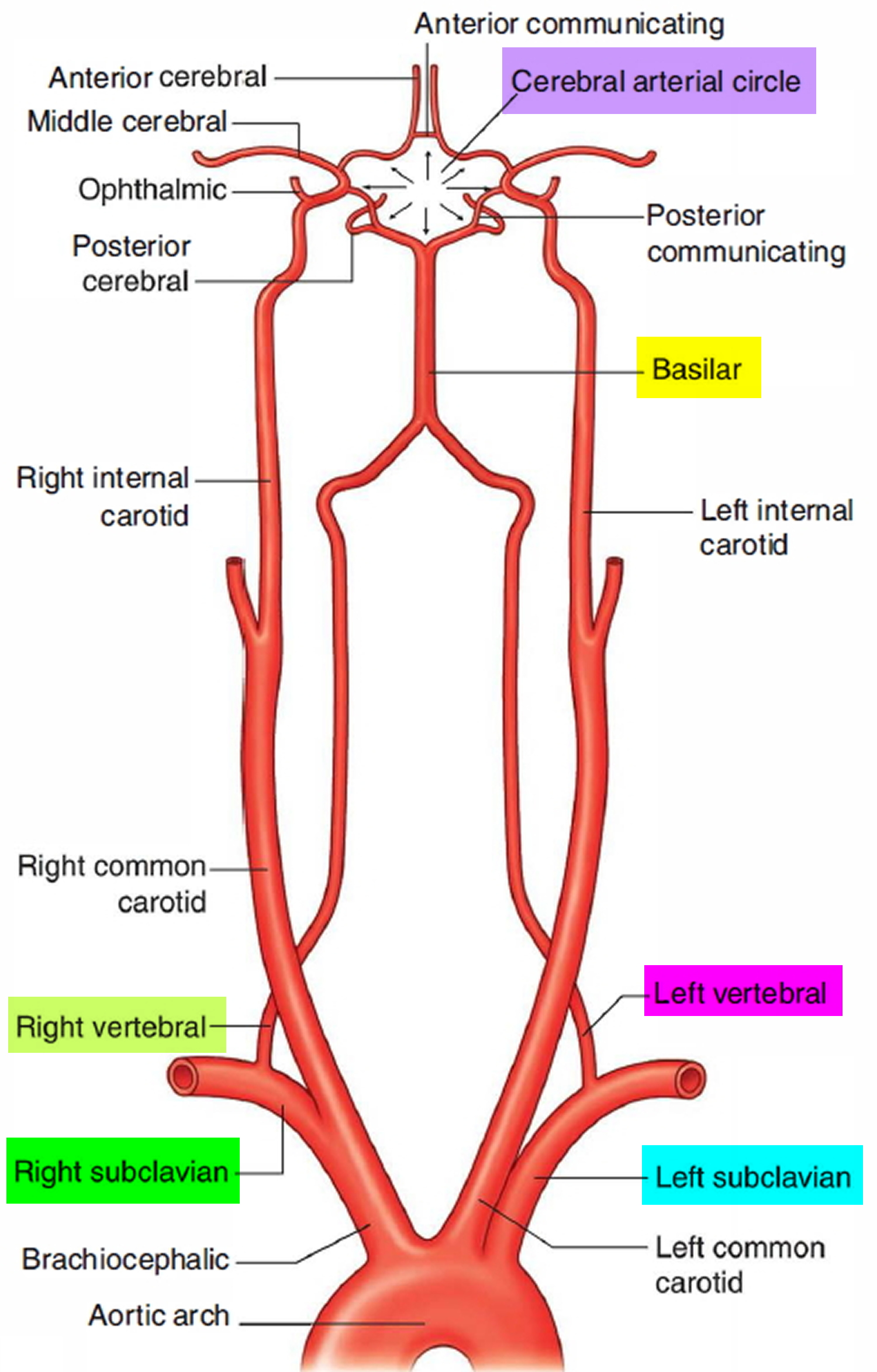
Left and Right Subclavian Artery Function, Branches, Stenosis
The vertebral column is drained by plexuses (networks) of veins. These venous plexuses are formed by spinal veins along the vertebral column, both inside and outside the vertebral canal. The plexuses include: Internal vertebral venous plexus (or the epidural venous plexus) External vertebral venous plexuses Anterior internal vertebral venous plexus